Original Article
Imaging
in Ocular Trauma Optimizing the Use of Ultrasound and Computerised Tomography
Sadaf
Imran, Saima Amin, M Imran Hameed Daula
Pak J Ophthalmol 2011, Vol. 27 No. 3
. . . . . . . . . . . . . . . .
. . . . . . . . . . . . . . . . . . . . . . . . . . . . . . . . . . . . . . . .
. . . . .. . .. . . . . . . . . . . . . . . . . . . . . . . . . . . . . . . . .
. . . .
See end of article for authors affiliations
..
.. Correspondence to: Sadaf Imran Resident Radiology Department of Radiology National Institute of Child Health, Karachi Submission of paper May 2011 Acceptance for publication September 2011
..
.. |
Purpose:
This
study was conducted to identify the role of ultrasound (US) and computerized
tomography (CT) scan in diagnosis of common ocular traumatic lesion. Material
and Methods: A cross sectional observational study was conducted over one
year period simultaneously at the Jinnah Postgraduate Medical Center (JPMC),
Karachi and the PNS SHIFA hospital, Karachi. Fifty patients with traumatic
ocular injuries who were referred by the ophthalmologist for radiological
evaluation were included in the study. Data regarding five common traumatic
lesions namely intraocular foreign body, vitreous hemorrhage, lens
dislocation, retinal detachment and choroidal detachment was analyzed. Results:
The
age of subjects included in this study ranged from 08 years to 60 years (mean
age was 28 ± 1 year). Ultrasound
was able to detect the pathologies in 93% of the patients when compared with
CT scan. CT scan showed higher accuracy compared to ultrasound in detecting
intraocular foreign body (25 patients out of which 24 cases were diagnosed by
ultrasound) vitreous hemorrhage (26 patients out of which 22 cases were
diagnosed on ultrasound) and lens dislocation (04 patients out of which 02
cases were diagnosed on ultrasound). However ultrasound showed higher
accuracy compared to CT scan in detecting retinal detachment (20 patients out
of which only 06 cases were diagnosed on CT scan) and choroidal detachment
(08 patients while CT was unable to detect any case of choroidal detachment). Conclusion: In the setting of acute ocular trauma CT scan
is more accurate in detecting intraocular foreign body, vitreous hemorrhage
and lens dislocation whereas ultrasound is superior in diagnosing retinal
detachment and choroidal detachment. Combined use of these imaging modalities
is recommended in diagnosis and management of post traumatic patients with
ocular injuries. |
Ocular trauma is common and may lead to vision threatening
pathologies. It has been estimated that world-wide ocular injuries are
responsible for blindness in approximately 1.6 million people, bilateral visual
impairment in 2.3 million and unilateral visual loss in about 19 million people
annually1. Approximately half of all patients who present to an eye
casualty department are suffering from ocular trauma2.
Ocular injuries are usually associated with injuries to head and
neck or limb structures and definitive diagnosis of ocular trauma in the acute
setting may be challenging. Missed ocular injuries in polytrauma patients may
lead to serious morbidity later on, necessitating the need of high index of
suspicion and employment of appropriate imaging techniques to accurately define
the extent and type of ocular injury.
Various imaging modalities exist to aid in the initial and
subsequent evaluation of trauma involving the eye and orbit. However, the best
modality for the initial evaluation of eye trauma remains indirect ophthalmoscopy.
In the first few hours after a severe injury, the first examiner can obtain
information with a level of detail that no other imaging method can provide.
Although the early view may not always be the best one, often the first look
into a traumatized eye is the only look. Direct visualization of intraocular
structures however may become difficult or impossible when the eye lids are
swollen after injury. The use of imaging modalities like ultrasonography and CT
scanning can be useful adjuncts in the management of such patients. Standard
roentgeno-graphy, computed tomography (CT), magnetic resonance imaging (MRI),
and ultrasonography have been employed in managing ocular trauma patients and
all have their strengths and weaknesses3-5. This study explores the
role of ultrasound and CT scanning in the management of ocular trauma.
MATERIAL AND METHODS
After appropriate technical and ethical approval from the relevant
review boards this cross sectional observational study was conducted simultaneously
at the Jinnah Postgraduate Medical Center (JPMC), Karachi and the PNS SHIFA
hospital, Karachi. Sampling technique was non-probability convenience. The
sample comprised fifty patients with traumatic ocular injuries who were
referred by the ophthalmologist for radiological evaluation. Data regarding
five common traumatic lesions namely intraocular foreign body, vitreous
hemorrhage, lens dislocation, retinal detachment and choroidal detachment were
analyzed. Period of study was 01 year from 31st January 2009 to 30th
January 2010. All the patients were examined by ultrasound in supine position
with linear high frequency transducer of 7.5 MHZ on single ultrasound machine
(GEVoluson 730), using closed eye technique with water soluble gel followed by
orbital CT scan. Spiral CT of patients was performed on Toshiba Asteion 16
slices CT scanner. Axial slices were obtained with 120 KV and 250MA. The CT
protocol included pitch of 1.0 slices thickness 1.0mm and reconstruction
interval 5.0 mm. Reformatted coronal and sagital images were also obtained. In
all selected patients the findings seen on Ultrasound and CT scan were
collected and proforma were filled for each patient.
RESULTS
In this study there were 34 (68%) male patients and 16 (32%) were
female patients. The mean age of patients in our study was 28± 1 year (SD
14.5).
After ultrasound all patients underwent CT scanning. Overall
ultrasound was able to detect the pathologies in 93% of the patients when
compared with CT scan results. Data analysis for five individual pathologies as
shown in tables 1 and 2 was carried out and it revealed that CT scan diagnosed
foreign body in 25 patients out of which 24 cases were correctly diagnosed on
Ultrasound. CT scan diagnosed vitreous hemorrhage in 26 patients out of which
22cases were correctly diagnosed on Ultrasound. CT scan diagnosed lens
dislocation in 04 patients out of which 02 cases were correctly diagnosed on
Ultrasound. Ultrasound diagnosed retinal detachment in 20 patients out of which
06 cases were correctly diagnosed on CT scan. Ultrasound diagnosed choroidal
detachment in 08 patients, none of these were picked up by CT scan.
Disease distribution include frequency of foreign body 25 (30%),
vitreous hemorrhage 26 (31%), retinal detachment 20 (24%), choroidal detachment
8(10%) and lens dislocation 4 (5%).Chi square test was applied to check
association between categorical variables (p<0.00).
DISCUSSION
Traumatic ocular emergencies can present in isolation or as part
of poly trauma. It is difficult to perform a physical examination on a severely
injured patient. The eye may be swollen shut or there may be intraocular
bleeding rendering fundoscopic examina-tion impossible. The patient may be
unable to cooperate or respond, making it difficult to evaluate for visual
acuity or ocular movement. Since these lesions can lead to exceptional
morbidity in the form of vision loss they warrant a high index of suspicion and
prompt and judicious use of imaging modalities to obtain an accurate diagnosis
and initiate appropriate management at an early stage.
Studies have shown that ultrasound and CT scan are highly accurate
in detection of ocular pathologies.6 Computed tomography is considered as having
advantage due to its ability of performing
multiplanar reformation, evaluating intraorbital structures with simultaneous
imaging the bony orbit for fractures and any herniations of orbital contents7.
Our study includes both children and adults with history of ocular
trauma. Mean age of patients was 28 ±1 year in our study while in study done by
Deramo et al it was 36 years7.
In this study ocular sonography was done using a single machine
(GE Voluson 730)and a standardized technique similar to that used by other
researchers (e.g. by Hoffman) using close eye technique with linear 7.5 10
MHz probe in sagittal and transverse plane8. Ultrasound was followed
by CT scanning done by similar protocol as described by Kazuhiro et al.9
Axial slices were obtained from above the orbit to below the orbit, the field
of view included the cavernous sinus and anterior brain stem with 120 KV and
100-160 MA, pitch of 1.5, slices thickness 1.0mm and reconstruction interval
1.0 mm. All CT scans done were on Toshiba Asteion 16 slice CT scanner.
Blaivas et al4 evaluated the accuracy of ultrasound for
the diagnosis of ocular pathology in patients with ocular trauma and findings
were confirmed by thin slices CT scanning. The results showed 100% sensitivity
and 97.2 % specificity. While in our study the sensitivity is 97.3% and
specificity is 94.4%.
Table 1: Patient gender distribution based on
pathology
Pathology |
Female |
Male |
Total patients |
Intraocular foreign body |
09 |
16 |
25 |
Vitreoushaemorrhage |
04 |
18 |
26 |
Ectopialentis |
02 |
02 |
4 |
Retinal detachment |
04 |
16 |
20 |
Choroidal detachment |
04 |
04 |
8 |
Table 2:
Ultrasound VS CT scan diagnostic breakdown based on different pathologies
Pathology |
Correctly diagnosed on Ultrasound |
Correctly diagnosed on CT scan |
Incorrectly diagnosed on Ultrasound |
Incorrectly diagnosed on CT scan |
Total |
Intraocular foreign body |
24 |
25 |
01 |
0 |
25 |
Vitreous haemorrhage |
22 |
26 |
4 |
0 |
26 |
Ectopialentis |
2 |
4 |
2 |
0 |
4 |
Retinal detachment |
20 |
6 |
0 |
14 |
20 |
Choroidal detachment |
8 |
0 |
0 |
8 |
8 |
Table 3: Sensitivity, specificity, PPV
(Positive Predictive Value), NPV (Negative Predictive Value) of ultrasound and
Ct Scan For Different Ocular Pathologies
Pathology |
Ultrasound |
Ct Scan |
||||||
Sensitivity % |
Specificity % |
PPV % |
NPV % |
Sensitivity % |
Specificity % |
PPV % |
NPV % |
|
Intraocular foreign body |
96 |
92.8 |
92.3 |
96.2 |
100 |
96.15 |
96.15 |
100 |
Vitreous haemorrhage |
84.6 |
96.5 |
95.6 |
87.5 |
96.29 |
92.3 |
92.85 |
96 |
Ectopialentis |
75 |
97.9 |
66.6 |
96 |
80 |
95.8 |
66.6 |
96 |
Retinal detachment |
95.2 |
90.9 |
86.9 |
96.7 |
30 |
95.6 |
75 |
75.8 |
Choroidal detachment |
80 |
97.7 |
88.8 |
91.6 |
0 |
98 |
0 |
86.2 |
Fig. 1: Oculr
ultrasound with linear probe
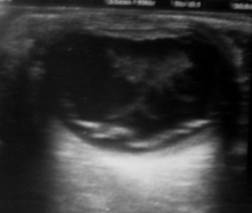
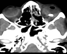
Fig. 2:
22-year male patient with vitreous hemorrhage, retinal detachment
and foreign body.
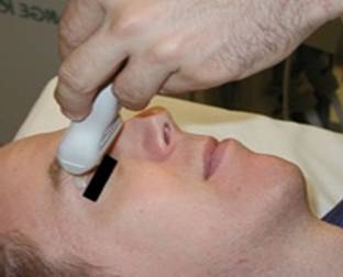
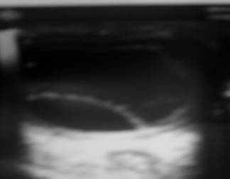
Fig. 3: 30-year female presented with history of
blunt trauma. Ultrasound image reveals retinal detachment.
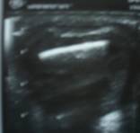
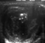
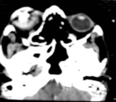
Fig. 4:
60-year-old male patient presented with oculartrauma during iron
working,Ultrasound and CT images show largeforeign body, vitreous hemorrhage
and retinal detachment.
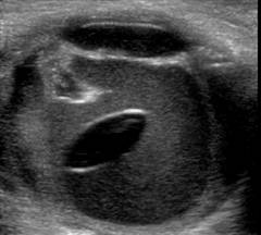
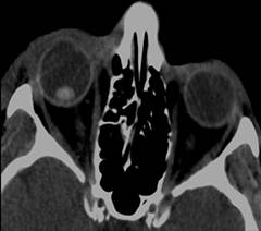
Fig. 5: 21 year old female patient presenting with
trauma. Imaging reveals Lens dislocation
Shiver et al3 stated the sensitivity of ultrasound for
detecting foreign body to be 87.5% and specificity to be 95.8 %. In our study
the sensitivity for foreign body detection was 96 % and specificity was 92.8%.
CT is considered the most sensitive method for detection of
intraocular foreign body reaching more than 95% detection rate10
while in our study the CT sensitivity for diagnosis of intraocular foreign body
reached 100%.
Ultrasonography is an excellent method to detect all kinds of
intraocular foreign bodies with an overall detection rate for metallic and non metallic
foreign body reaching 93% stated by Khan & Khan et al11.
In our study the sensitivity of ultrasound for the diagnosis of
retinal detachment is 95.2% & specificity is 90.9% in comparison with
Dhakshina et al the sensitivity is 92.3 % and specificity is 100%12 hence
retinal detachment is well demonstrated by ultrasound as well as sometimes by
CT as a 'V' or a 'sunset sign13.
In our study the sensitivity of ultrasound for the diagnosis of
vitreous hemorrhage is 84.6% and specificity is 96.5 % in comparison with S.
Kim S. Lee et al sensitivity is 73% and specificity 90%14.
Gilbert et al stated that sensitivity of CT for the diagnosis of
lens dislocation is 100% and specificity is 96% 15while in our study
the sensitivity of CT is 80 % and specificity is 95.8%. Dislocation of lens
into opaque media is a perfect indication for ultrasound. The abnormally placed
lens is easily detected because of its shape and strong reflectivity. Munk et
al (1991) demonstrated lens fragmentation with individual fragments distinctly
discernible on ultrasound16.
The slight difference of results in my study in comparison with
other studies was possibly due to incorporation of patients and operator
dependency of ultrasound and CT scan done with 3-5 mm slice thickness due to
patient load and time limitation rather than 1.0mm, which is used in other
studies9.
Ultrasound provides good visualization of ocular anatomy that
allows evaluation of intraocular foreign body and related lesions such as
vitreous hemorrhage and retinal detachment17.Ultrasound is
inexpensive and readily available in most Radiology departments. On the other
hand it is operator dependent technique. The examination of the globe is
exhaustive and patient is asked to perform ocular movements to find the exact
ultrasound incidence angle to visualize the foreign body18, however
ultrasound is useful in detecting small, nonmetallic posteriorly located
foreign bodies that may not be detected by other methods19,20.
CT is accurate in detecting and localizing intraocular, metallic,
glass and stone foreign bodies,21 CT imaging offers short
examination time and has the ability to obtain diagnostically useful coronal
and sagittal reconstruction images21 on the other hand there is
radiation dose delivered to the lens. In the presence of significant facial
trauma it is very difficult to determine the cause of decreased visual acuity.
Significant vitreous hemorrhages, intraocular foreign bodies, chorioretinal
detachment, lens dislocation and others all result in visual loss and require
imaging for diagnosis.
This study shows that ultrasound has high sensitivity and
specificity in diagnosing traumatic ocular diseases and is superior to CT scan
in diagnosing retinal detachment and choroidal detachment while CT scan detects
foreign body, vitreous hemorrhage and lens dislocation more accurately than
ultrasound.
The results of this study support the combined use of ultrasound
and CT scan imaging in managing patients with traumatic ocular injuries who are
referred for radiological evaluation. However keeping in view the common
availability, cost effectiveness and acceptably high sensitivity and
specificity of ultrasound in detecting ocular traumatic pathologies the authors
strongly propose liberal use of ultrasound in managing these patients. The
importance of incorporating ocular ultrasound training for all radiologists,
ophthalmologists and emergency department physicians cannot be overemphasized.
Authors affiliation
Dr.
Sadaf Imran
Resident
Radiology
Department
of Radiology
National Institute of Child
Health, Karachi
Dr.
Saima Amin
Assistant
Professor
Department
of Radiology
Jinnah Postgraduate Medical
Centre, Karachi
Dr. M
Imran Hameed Daula
Department of Surgery
PNS SHIFA Hospital, Karachi
REFERENCE
1.
Negrel AD, Thylefors B. The
global impact of eye injuries. Ophthalmic Epidemiol. 1998; 5: 143-69.
2.
Chiapella AP, Rosenthal AR. 1
year in an eye casualty clinic. Br J Ophthalmol. 1985; 69: 865-70.
3.
Stephen A. Shiver, Mathew Lyon,
Micheal Blaivas. Detection of Metallic Ocular foreign Bodies with Handheld
Sonography in a porcine Model. J ultrasound Med. 2005; 24: 1341-6.
4.
Blaivas M. Bedside emergency
department ultrasonography in the evaluation of ocular pathology. AcadEmera
Med. 2000; 7: 947-50.
5.
Bord SP, Linden J.
Trauma to the globe and orbit. Emerg Med Clin North Am. 2008; 26: 97123.
6.
Blaivas M Theodor D, Sierzenski P. A study of bedside ocular ultrasonography in the emergency
department. AcadEmer Med. 2002; 9: 791-9.
7.
DeramoVa, Shah Gk, Baumal CR, et al. Role of ultrasound biomicroscopy in ocular trauma. Trans Am
Ophthamol Soc. 1998; 96: 355-65.
8.
Ultrasound guide for
emergency physician, an introduction, Beatrice Hoffmann, MD, PhD, RDMS.
9.
Katada K, Kauczor HU, Schuzer J, et al. Multidetector CT protocol-developed for Toshiba scanner, Spring
2005.
10.
NovellineRA, Liebig T, Jordan J, et al. Computed tomography of ocular trauma. Emerg Radiol. 1994; 1:
5667.
11.
Khan BS, Khan MD; A
Review of 100 cases of Ectopialentis. Presentation, Management and visual
prognosis, Pak J of Ophthalmol: 2002; 18: 3-9.
12.
Ganeshan DM. Probing into Retinal
detachment ultrasound is eminently useful as diagnostic tool. 2008; 31,.
13.
Pieramici DJ. Vitreo retinal
trauma.OphthalmolClin North Am. 2002; 15: 225-4.
14.
Kim S. Lee comparison of
ultrasound & intraoperative findings in patients with vitreous hemorrhage,
invest Ophthalmol Vis Sci. 2005; 46: 5436.
15.
CE. Gilbard CRO 15.1, CT & US in lens dislocation & IOF Bs
18.5: 118.
16.
Munk PL, Vellet AD, Levin M, et al. Sonography of the eye. Am J Roentgenol. 1991; 157: 1079-86.
17.
Berges O. Orbital
ultrasonography: Principles and technique. In: Newton TH, ed. Radiology of the
Eye and Orbit. New York, NY: Raven Press. 1990: 6.16.20.
18.
DeramoVa, Shah Gk, Baumal CR, et al. Ultrasound biomicroscopy as a tool for Detecting and localizing
occult foreign bodies after ocular trauma Ophthalmology. 1999; 106: 301-5.
19.
DeramoVa, Shah Gk, Baumal CR, et al. Role of ultrasound biomicroscopy in ocular trauma. Trans Am
Ophthamol Soc. 1998; 96: 355-65.
20.
Lakitas A, Prokesch R, Scholda C, et al. Orbital helical computed tomography in the diagnosis and
management of eye trauma Ophthalmology. 1999; 106: 2330-5.
21.
Dass AB, Ferrone PJ, Chu YR, et al. Sensitivity of spiral computed tomography scanning for detecting
intraocular foreign bodies. Ophthalmology. 2001; 108: 23268.